Introduction: Deep Relief for Your Back – Understanding Lumbar Decompression
Back pain, especially in the lower back, can be a debilitating experience that significantly affects quality of life. When conservative treatments do not provide the relief you hoped for, lumbar decompression emerges as a group of surgical procedures designed with one clear aim: to relieve the pressure on the nerve roots or the spinal cord in the lower back. This article looks at what lumbar decompression involves, why it is done, the different types of procedure available, what to expect during the process and, most importantly, how it can help patients find lasting relief and recover their wellbeing.
It is essential to understand that lumbar decompression does not refer to a single surgical technique. It is an umbrella term for several strategies with a common goal: freeing the compressed nerve structures. In fact, lumbar decompression is considered the cornerstone of the surgical treatment of degenerative conditions of the lumbar spine.1 The essential purpose of these procedures is to create more room inside the spinal canal, relieving the pressure on the spinal cord and nerves.2 The need for lumbar decompression usually arises from an underlying condition that compresses the nerves, and lumbar spinal stenosis is one of the most common.
The Root of the Problem: Why Might You Need Lumbar Decompression? (Lumbar Spinal Stenosis)
To understand lumbar decompression, it helps first to know the main condition it aims to treat: lumbar spinal stenosis.
What is lumbar spinal stenosis?
Lumbar spinal stenosis is, in simple terms, a narrowing of the spinal canal through which the spinal cord (or, more precisely, the cauda equina in the lumbar region) and the nerve roots running down to the legs pass.2 It is particularly common in people over 50 or 60, and its prevalence tends to increase with age, becoming more noticeable from the age of 40.2 In fact, an overall prevalence of around 11% of the population has been estimated, with an average age of those affected of about 62.4
Lumbar spinal stenosis is a common condition, especially in people over 60, in which the space for the nerves in the spine becomes reduced.
From an anatomical point of view, the normal anteroposterior (AP) diameter of the lumbar spinal canal ranges from 22 to 25 mm. Stenosis is described as relative when this diameter falls to 10-12 mm, a situation that is often without symptoms. Absolute stenosis, with an AP diameter below 10 mm, usually causes obvious symptoms.4 It is important to stress that not everyone with imaging evidence of stenosis will experience disabling symptoms; the treatment decision is based fundamentally on the patient’s clinical picture.
Common causes: natural wear and tear and other culprits
The most frequent cause of lumbar stenosis is the degenerative process associated with ageing and arthritis.2 Over time, several changes can occur in the spine:
- Bone spurs (osteophytes): Bony growths that can encroach on the space of the canal.
- Degeneration of the intervertebral discs: The discs lose height and water content, and may bulge or herniate into the canal.
- Thickening of the ligaments: Particularly the ligamentum flavum, which can hypertrophy and reduce the available space.
- Osteoarthritis of the facet joints: The small joints that connect the vertebrae can become inflamed and enlarged. 3
Less common causes include significant disc herniations that by themselves cause narrowing2, spondylolisthesis (a slip of one vertebra over another; you can find more information in our article on lumbar spondylolisthesis)6, trauma, and, more rarely, tumours or congenital conditions in which the spinal canal is narrow from birth.2
Symptoms you should not ignore
The symptoms of lumbar stenosis can vary, but some are very characteristic:
- Neurogenic claudication: This is the classic symptom. It shows up as pain, cramping, heaviness or weakness in one or both legs and/or buttocks that appears or gets worse when walking a certain distance or standing for long periods. Typically, these symptoms improve or disappear on sitting down or leaning the trunk forward.5
- “Shopping trolley sign”: Many patients find that their symptoms ease when they lean forward, for example when resting on a shopping trolley. This posture temporarily increases the space in the lumbar canal, relieving pressure on the nerves.4 Recognising this sign can be an important clue for patients.
- Low back pain: It may be present, and is often chronic or long-standing, although not all patients with stenosis experience it prominently.6
- Sciatica (radicular pain): Pain that radiates from the lower back into the leg, following the path of a compressed nerve. It is often described as sharp, burning or like an electric shock. It may come with numbness, tingling (paraesthesia) or a “pins and needles” sensation in the leg or foot.6 Disc herniations are also a common cause of this symptom.
- Leg weakness: There may be difficulty lifting the foot (foot drop) or weakness in other muscle groups of the legs.7
- Cauda equina syndrome: In rare but serious cases of severe stenosis, massive compression of the nerves of the cauda equina can occur, leading to symptoms such as loss of bladder or bowel control, numbness in the genital or perianal area (“saddle anaesthesia”) and severe leg weakness. This is a medical emergency that requires urgent surgery.3
The road to diagnosis: how does your doctor know it is stenosis?
The diagnosis of lumbar spinal stenosis is based on a combination of the clinical history, the physical examination and imaging tests:
- Detailed clinical history: The doctor will ask in depth about your symptoms: when they started, what triggers them, what relieves them, how they affect daily activities, and so on.8
- Physical and neurological examination: Muscle strength in the legs, sensitivity to touch and pain, and tendon reflexes will be assessed. The doctor may also ask you to walk or adopt certain postures to see whether the symptoms are reproduced.6
- Imaging tests:
- X-rays: They can show indirect signs of degeneration such as loss of disc height, the presence of bone spurs or the alignment of the vertebrae (for example, in spondylolisthesis).6
- Magnetic resonance imaging (MRI): This is the most useful imaging test and is often considered the test of choice for diagnosing lumbar stenosis. It provides detailed images of soft tissues, including the intervertebral discs, ligaments and nerve structures, allowing direct visualisation of the narrowing of the canal and the compression of the nerves.8
- Computed tomography (CT): It offers excellent visualisation of bony structures and can be useful for accurately measuring the dimensions of the spinal canal, or for assessing bony detail in patients who cannot have an MRI.8
It is important to remember that imaging findings must always be correlated with the patient’s symptoms. Many people can have signs of stenosis on an MRI without experiencing significant symptoms. The treatment decision, especially when it comes to surgery, is based on the impact these symptoms have on the patient’s daily life.
Lumbar Decompression Surgery: Making Room for Your Wellbeing
The primary goal of lumbar decompression surgery is to relieve the pressure on the nerve structures (whether individual nerve roots or the cauda equina) inside the lumbar spinal canal. By creating more room, the aim is to reduce pain (especially pain that radiates into the legs), numbness and muscle weakness and, ultimately, to improve the patient’s functional capacity and quality of life.1
When is surgery considered?
The decision to undergo lumbar decompression is not taken lightly. It is generally considered an option when:
- Conservative treatment has failed: This includes measures such as medication (painkillers, anti-inflammatories), physiotherapy, activity modification and, in some cases, epidural corticosteroid injections. If these treatments, applied for a reasonable period, do not achieve satisfactory relief of symptoms, surgery may be the next step.9
- Symptoms are severe, progressive or disabling: When pain, weakness or numbness significantly limit the patient’s ability to walk, stand, work or carry out daily activities, the surgical option is considered.9 Pain is often the deciding factor in this choice.10
- There are significant neurological deficits: Progressive muscle weakness in the legs, or a “foot drop” (difficulty lifting the tip of the foot), are clear indications to consider surgery in order to avoid permanent nerve damage.9
- Cauda equina syndrome: As mentioned above, this is an emergency that requires immediate lumbar decompression to prevent irreversible damage to nerve function, including bladder and bowel control.3
A 10-year prospective study on the management of lumbar stenosis found that, although an initial conservative approach is advisable for many patients, surgical treatment offered more favourable results in the long term. It is worth noting that patients in the conservative group whose outcome was not satisfactory could be treated surgically later with a good prognosis.10 This underlines that surgery is a valuable tool when properly indicated, and that the decision is made by considering the overall impact of the symptoms on the patient’s life. Surgery mainly aims to relieve pain radiating into the legs (sciatica, claudication), although axial low back pain (pain in the back itself) may persist if its origin is osteoarthritis of the joints, because decompression surgery does not cure the underlying arthritis.1
A Journey Through the Techniques: Types of Lumbar Decompression Surgery
Lumbar decompression surgery has evolved remarkably. Surgical techniques have moved on from more extensive procedures to minimally invasive approaches, with the aim of reducing surgical trauma, preserving as much healthy spinal structure as possible and speeding up the patient’s recovery. The most common types of lumbar decompression surgery are described below:
Table 1: Quick Comparison: Common Types of Lumbar Decompression Surgery
| Surgical technique | Brief description of the procedure | Level of invasiveness (general) | Main indication/focus |
| Laminectomy | Removal of the lamina (the back part of the vertebra) to widen the spinal canal. | Traditional/open | Extensive stenosis, often at several levels. |
| Laminotomy/foraminotomy | Removal of a portion of the lamina (laminotomy) or widening of the foramen (foraminotomy). | Less invasive/open or MIS | Localised nerve compression, foraminal or lateral recess stenosis. |
| Discectomy | Removal of the herniated or bulging portion of the disc that is compressing a nerve. | Open or MIS | Disc herniation contributing to nerve compression (may or may not be associated with stenosis). |
| Microdecompression | Laminectomy, laminotomy or discectomy performed with a microscope and small incisions. | Minimally invasive (MIS) | Similar to open techniques but with less tissue trauma. |
| MILD® procedure | Percutaneous, image-guided removal of excess thickened ligamentum flavum. | Percutaneous/MIS | Central canal stenosis due to hypertrophy of the ligamentum flavum. |
| Endoscopic surgery | Use of an endoscope (camera and small instruments) through very small incisions. | Minimally invasive (MIS) | Disc herniations, localised central canal or foraminal stenosis, with minimal tissue trauma. |
The evolution: from traditional laminectomy to minimal invasion
Historically, lumbar decompression was performed with more open techniques which, although effective at freeing the nerves, could involve greater disruption of the muscles and other structures of the back. Over time, the development of specialised instruments and advanced imaging techniques has made it possible to carry out these procedures in a far less aggressive way.
Laminectomy: the classic approach
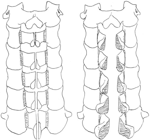
A laminectomy consists of removing the lamina, the bony part at the back of the vertebra that covers the spinal canal.3 Removing the lamina increases the diameter of the canal, relieving pressure on the nerve structures. It is a well-established technique and can be very effective, especially in cases of severe stenosis or stenosis affecting several vertebral levels.8
Although it is more invasive than modern techniques, it remains a valid option. A historical concern was possible postoperative vertebral instability, especially if a large part of the facet joints was removed. However, studies indicate that instability is relatively rare if these joints are adequately preserved.13 If instability is anticipated or created, laminectomy can be combined with a spinal fusion.9
Laminotomy and foraminotomy: precision to relieve pressure
These procedures are more selective than laminectomy:
- Laminotomy: This involves creating a small opening or window in the lamina, removing only a portion of bone to reach the spinal canal and decompress the affected nerve. It is less extensive than a laminectomy.15
- Foraminotomy: It focuses on enlarging the intervertebral foramen, the side opening through which each nerve root leaves the spine. It is done when the narrowing is located at this exit, often because of bone spurs or a foraminal disc herniation.15 Both techniques can be performed on their own or combined with other procedures, such as discectomy, and often benefit from the use of a microscope (microdecompression).17

Discectomy: when a disc is part of the problem
A discectomy is the surgical removal of the part of an intervertebral disc that has herniated or bulged and is compressing a nerve root or the spinal cord.15 Although discectomy is commonly associated with the treatment of disc herniation, it is also a form of lumbar decompression when the herniation contributes significantly to stenosis of the canal or the foramen.
Modern advances: less invasive options
The aim of minimally invasive (MIS) techniques is to achieve the same decompression goals as open surgery, but with less damage to the surrounding muscles and tissues, which ideally means less postoperative pain, a shorter hospital stay and faster recovery.
- Microdecompression and the MILD® procedure: specific solutions.
- Microdecompression uses a surgical microscope to magnify the field of view, allowing the surgeon to perform the decompression (whether a laminectomy, laminotomy or discectomy) through much smaller incisions.
- The MILD® procedure (Minimally Invasive Lumbar Decompression) is an even less invasive percutaneous (through the skin) technique, guided by imaging (fluoroscopy). It is specifically designed to treat central canal stenosis caused by hypertrophy (thickening) of the ligamentum flavum.20 It is carried out with small tools that remove portions of the thickened ligament, thereby increasing the space for the nerves.
- Indications: Patients with neurogenic claudication whose symptoms are mainly due to hypertrophy of the ligamentum flavum and who have not improved with conservative treatment.20
- Efficacy and safety: Multiple studies, including systematic reviews and meta-analyses, have shown that MILD® significantly reduces pain (measured with the Visual Analogue Scale, VAS) and disability (measured with the Oswestry Disability Index, ODI), with results maintained for up to two years. Its efficacy has been reported to be superior to epidural steroid injections, with a comparable safety profile and a low rate of adverse events.20
The MILD® procedure is a safe and effective minimally invasive option for specific patients with lumbar stenosis caused by thickening of the ligamentum flavum.
- Endoscopic spine surgery: cutting-edge technology for faster recovery.
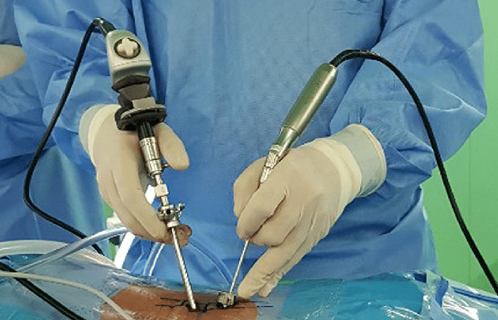
Endoscopic spine surgery is one of the most significant advances in minimally invasive lumbar decompression. It uses an endoscope, a thin tube with a high-definition camera and a light at its tip, which is introduced through a very small incision (usually under 1 cm). Through working channels in the endoscope, the surgeon introduces tiny instruments to carry out the decompression.23
- Advantages: The main benefit is minimal disruption of the muscles and soft tissues of the back. This leads to less blood loss during surgery, less postoperative pain, a shorter hospital stay (it is often a day-case procedure or needs one night in hospital) and a faster return to daily activities.23 In addition, by better preserving bone and ligament structures, it may reduce the risk of postoperative instability.23
- Applications: Endoscopic surgery can be used to treat disc herniations (endoscopic discectomy), central canal stenosis and foraminal stenosis (endoscopic foraminotomy), using transforaminal approaches (through the foramen) or interlaminar approaches (between the laminae).23
- Considerations: It requires specialised training and a learning curve on the part of the surgeon to master the technique.23 Not all lumbar conditions can be treated endoscopically, so patient selection is crucial.
The choice between these techniques depends on multiple factors, including the exact cause and location of the nerve compression, the number of vertebral levels affected, the presence of associated instability, the patient’s general health and, fundamentally, the surgeon’s experience and preference with each technique.
The role of spinal fusion: is it always necessary with decompression?
Spinal fusion (or arthrodesis) is a surgical procedure that permanently joins two or more vertebrae to eliminate movement between them and stabilise that segment of the spine. It is not a routine component of every lumbar decompression.
Fusion is considered together with decompression in specific situations25:
- Pre-existing instability: If the patient already has instability in the segment to be decompressed, as in the case of significant spondylolisthesis (vertebral slippage).
- Iatrogenic instability: If the decompression needed to adequately free the nerves involves removing a significant portion of the facet joints or other stabilising structures, which could create instability.
- Associated deformity: In cases of degenerative scoliosis or other deformities that require correction and stabilisation.
Modern lumbar decompression techniques, especially minimally invasive ones, aim precisely to preserve the stabilising structures as far as possible so that fusion is not needed.13 When instability is a problem, there are various fusion techniques, some of them also minimally invasive, such as TLIF (Transforaminal Lumbar Interbody Fusion). In some selected cases of mild instability, or to prevent it, devices such as interspinous spacers can be considered.16
After Lumbar Decompression Surgery: Expectations and Recovery
Undergoing lumbar decompression surgery is an important step, and knowing what to expect afterwards is essential for a successful recovery and for setting realistic expectations.
Expected results: pain relief and better quality of life
The main goal of lumbar decompression is to relieve radicular pain, that is, pain that radiates down the legs because of nerve compression. Most patients experience a significant improvement in this type of pain. Purely lumbar pain (pain in the back) may improve too, but often to a lesser extent, especially if it is related to underlying osteoarthritis that decompression itself does not correct.
As well as pain relief, an improvement is expected in the ability to walk, stand and carry out activities of daily living that were previously limited by symptoms. Studies using rating scales such as the Visual Analogue Scale (VAS) for pain and the Oswestry Disability Index (ODI) for function consistently show improvement after surgery.
A long-term follow-up study found that 73% of patients achieve a “minimal clinically important difference” (MCID) two years after surgery, and these benefits tend to be maintained over time.
Studies show that lumbar decompression surgery is successful in improving leg pain in most patients, with benefits maintained in the long term.
Factors that influence your recovery
Every patient’s recovery is unique, but certain factors can influence the process and the results:
- Type of surgery: Minimally invasive techniques (such as microdecompression or endoscopic surgery) are generally associated with shorter recovery times, less postoperative pain and a faster return to activities compared with traditional open surgery.23
- General health and age: Although advanced age on its own is not a contraindication (laminectomy has been shown to be safe even in people over 80 14), the presence of other illnesses (comorbidities) can influence recovery.
- Psychosocial factors: A higher level of education, optimistic preoperative expectations and good emotional health have been observed to correlate with better postoperative results.29 The patient’s attitude and commitment to recovery are therefore very important.
- Smoking: Smoking can negatively affect healing and has been associated with less relief of leg pain after surgery.29 Stopping smoking before surgery is strongly recommended.
- Specific diagnosis: In some studies, patients operated on for a disc herniation as the cause of the compression have reported greater relief of back pain and functional improvement compared with other causes of stenosis.29
- Primary versus revision surgery: Generally, a first lumbar decompression surgery tends to have more predictable and effective results than revision surgery (when a previously operated area is operated on again).1
The road back to normal: postoperative care and rehabilitation
The postoperative phase is crucial to secure the benefits of surgery.
Table 2: Preparing for Surgery and Recovery: Key Points
| Phase | Key actions/considerations |
| Before surgery | Discuss everything thoroughly with your surgeon and make a list of questions. Optimise your health: stop smoking and control your weight if possible.6 Prepare your home to make recovery easier (for example, remove obstacles and have help available).18 |
| Immediately afterwards (hospital) | Pain management with prescribed medication. Early mobilisation assisted by physiotherapists or nursing staff. Learning the “logroll” technique for moving around. Care of the surgical wound.9 |
| First weeks at home | Follow the movement restrictions strictly: do not bend at the waist, do not twist the trunk, do not lift heavy objects.12 Wear the brace if one has been prescribed. Take short, progressive walks. Keep an eye on the wound and contact your doctor if there are signs of infection (redness, discharge, fever).33 Many of the movement recommendations given after surgery with implants also apply here. |
| Long-term recovery | Physiotherapy and a personalised exercise programme as indicated by your doctor. The aim is to strengthen the back and core muscles and to improve flexibility and posture.9 Gradual return to work and recreational activities. Maintain a healthy lifestyle (suitable weight, regular exercise) to protect your back in the future.6 |
- Hospital stay: It is usually short. For a simple laminectomy, it can be 1 to 2 days.9 Endoscopic techniques may even allow discharge on the same day or the following day.24
- Pain management: Painkillers will be prescribed to control pain at the incision site and residual muscle pain.9
- Initial restrictions (spinal precautions): During the first few weeks, it is vital to protect the spine. This means:
- Do not bend at the waist (lumbar flexion): Avoid stooping or leaning forward.
- Do not twist the trunk (torsion): Move the body as a single block.
- Do not lift heavy objects: The limit is generally under 4-5 kilograms.
- The “logroll” technique is taught for getting out of bed and moving around, keeping the back straight like a log.12
- Use of an orthosis (brace): It is not always necessary, but the surgeon may prescribe it to provide support and limit movement during the early phases of recovery, especially if a fusion has been performed or if there are concerns about stability.12
- Rehabilitation and physiotherapy: Physiotherapy plays an important role in recovery. A personalised exercise programme will help restore the strength of the back and abdominal muscles (the “core”), improve flexibility and posture, and relearn safe movement patterns.9 Core strengthening is essential, and stretching, such as hamstring stretches, is also beneficial.35 Interestingly, a randomised controlled trial investigated the effect of postoperative rehabilitation and found that a 12-week supervised physiotherapy programme offered no significant advantage in terms of pain or disability at 2 years, compared with simply advising patients to stay active by doing the physical activities they enjoyed most.36 This does not invalidate physiotherapy, which can be crucial for learning the right exercises and progressing safely, but it does highlight the importance of general physical activity and of empowering the patient in their own recovery.
- Return to activities: This is a gradual process. Returning to work will depend on the type of surgery and the nature of the job. Office activities can be resumed within a few weeks, while jobs that involve physical effort will take longer, possibly several months.9
Key Considerations: Risks and Possible Complications of Lumbar Decompression
Although lumbar decompression surgery is generally safe and effective when properly indicated, like any surgical procedure it carries certain risks and the possibility of complications. It is essential that patients are well informed so that they can make a conscious decision together with their surgeon.
Risks inherent to any spine surgery:
These are general risks associated with most surgical interventions on the spine:
- Reactions to anaesthesia: Most lumbar decompression operations are performed under general anaesthesia, which has its own risks, although these are low in healthy patients.18
- Bleeding: It can occur during or after surgery and, on rare occasions, can be significant.9
- Infection: It can be superficial (in the wound) or deep (affecting the bones or the disc space). The risk is generally low (1-3% or less), and lower still with minimally invasive techniques. Preventive antibiotics are given.9
- Blood clots (deep vein thrombosis, DVT): Clots can form in the veins of the legs, especially with reduced mobility. If a clot travels to the lungs (pulmonary embolism, PE), it can be serious. Preventive measures are taken.9
- Nerve injury: There is a small risk of damaging the nerve roots or the dura mater (the membrane that covers the nerves). This could cause persistent pain, numbness, weakness or, in very rare cases, paralysis. Intraoperative neurophysiological monitoring helps to minimise this risk.9
- Cerebrospinal fluid (CSF) leak: If the dura mater is accidentally torn, CSF can leak. This is often repaired during the same operation, but it can cause postoperative headaches and, rarely, require another operation.9
- General medical complications: Such as heart or lung problems, especially in patients with pre-existing conditions or of advanced age.12
Complications specific to lumbar decompression:
- Persistent or recurrent pain: Not all patients experience complete pain relief, or the pain may return over time. This can be due to multiple factors, including progression of degeneration at other levels or the formation of scar tissue.9
- Spinal instability: If the decompression requires removing a significant part of the facet joints or other structures that give stability to the spine, instability may develop. This could require a second operation to perform a spinal fusion.9 Modern techniques aim to minimise this risk.
- Disc re-herniation: If a discectomy was performed as part of the decompression, there is a possibility that the disc may herniate again at the same site or at an adjacent level.40
- Epidural fibrosis: This is the formation of scar tissue around the nerve roots after surgery. In some cases, this scar tissue can cause chronic pain.
Failed back surgery syndrome (FBSS) or post-laminectomy syndrome (PLS): a reality to be aware of
This is a term used to describe a situation in which low back pain, with or without pain radiating into the legs, persists or even worsens after spine surgery, without a clearly identifiable or easily correctable cause.39
- Prevalence: Estimates vary, but some studies suggest that a not insignificant percentage of patients (between 10% and 40%, and even up to 60% in specific series) may not experience the expected benefits of surgery or may develop chronic postoperative pain.39
- Impact: FBSS can be very frustrating and debilitating, causing intense chronic pain, a significant reduction in quality of life, functional disability and emotional problems such as anxiety and depression.39 Its management is complex and often requires a multidisciplinary approach.
It is crucial that patients discuss all these risks openly with their surgeon. Good patient selection, meticulous surgical technique and appropriate postoperative management are the keys to minimising complications.
Conclusion: Making an Informed Decision About Your Lumbar Decompression
Lumbar decompression surgery is a valuable and often very effective treatment option for patients suffering from symptomatic lumbar spinal stenosis and other conditions that cause nerve compression. The main benefits, such as relief of leg pain (neurogenic claudication, sciatica) and improved functional capacity, can make a substantial difference to the quality of life of those affected.
As described, the decision to proceed with a lumbar decompression is generally taken after conservative treatments have failed to provide the desired relief, and when symptoms are severe enough to have a negative impact on daily activities. The evolution of surgical techniques, from traditional approaches to minimally invasive procedures such as microdecompression, MILD® or endoscopic surgery, now offers a range of options that seek to maximise results and minimise the impact of the intervention.
It is essential that every patient has a thorough and open discussion with their spine specialist. This conversation should cover in detail the individual symptoms, the different treatment options (both conservative and surgical), the particulars of each proposed surgical technique, the benefits that can realistically be expected and the potential risks inherent in each procedure. Understanding these aspects allows patients to take an active part in the decision-making process, a partnership that is key to a satisfactory outcome.
While lumbar decompression can offer a new chance of a more active, pain-free life, information and careful consideration are the best allies. With the right knowledge and the support of an experienced medical team, patients can make the decision that best suits their needs and aspirations for wellbeing.
If you live outside Spain and would like Dr. Ferrández to review your case, you can book a video consultation or a written assessment on our booking page.
Further Resources and Links of Interest
For patients who want to explore related topics in more depth, we suggest the following articles on this site:
- Back stretching: the benefits
- The perils of stretching
- Lumbar spondylolisthesis: types, grades and treatment options
- Spinal stenosis
- Lumbar over-the-top decompression
External links:
For scientific research and more medical information (it is advisable to search with the help of a healthcare professional to interpret the studies):
- PubMed (database of biomedical literature): https://pubmed.ncbi.nlm.nih.gov/ (search for terms such as “lumbar decompression”, “spinal stenosis surgery”, “laminectomy outcomes”).
- Mayo Clinic: information on spinal stenosis 2 and laminectomy.9
- MedlinePlus (US National Library of Medicine): information on laminectomy 18 and foraminotomy.17
References
- Postural hygiene measures after lumbar spine surgery with implants (Dr. Ferrández’s own guidance article), accessed 11 May 2025
- Patient-Reported Outcomes Following Lumbar Decompression …, accessed 13 May 2025, https://pubmed.ncbi.nlm.nih.gov/32875849/
- Spinal stenosis – Symptoms and causes – Mayo Clinic, accessed 13 May 2025, https://www.mayoclinic.org/diseases-conditions/spinal-stenosis/symptoms-causes/syc-20352961
- Lumbar Spinal Stenosis – OrthoInfo – AAOS, accessed 13 May 2025, https://orthoinfo.aaos.org/en/diseases–conditions/lumbar-spinal-stenosis/
- Lumbar spinal stenosis | Diagnosis and treatment – Physiotutors (in Spanish), accessed 13 May 2025, https://www.physiotutors.com/es/conditions/lumbar-spinal-stenosis/
- Spinal decompression surgery (Dr. Ferrández’s own article, in Spanish), accessed 13 May 2025
- Lumbar Spinal Stenosis – StatPearls – NCBI Bookshelf, accessed 13 May 2025, https://www.ncbi.nlm.nih.gov/books/NBK531493/
- Symptoms of lumbar spinal stenosis – Spine-health (in Spanish), accessed 13 May 2025, https://www.spine-health.com/es/condiciones/estenosis-raquidea/sintomas-estenosis-espinal-lumbar
- Degenerative lumbar spinal stenosis – SciELO Cuba (in Spanish), accessed 13 May 2025, http://scielo.sld.cu/scielo.php?script=sci_arttext&pid=S1025-02552013000400009
- Laminectomy – Mayo Clinic, accessed 13 May 2025, https://www.mayoclinic.org/tests-procedures/laminectomy/about/pac-20394533
- Lumbar spinal stenosis: conservative or surgical management?: A …, accessed 13 May 2025, https://pubmed.ncbi.nlm.nih.gov/10828926/
- Treatments for spinal stenosis | Spine specialist – New York Spine Specialist (in Spanish), accessed 13 May 2025, https://newyorkspinespecialist.com/es/es-types-of-pain/estenosis-espinal/
- www.brighamandwomens.org, accessed 13 May 2025, https://www.brighamandwomens.org/assets/BWH/patients-and-families/rehabilitation-services/pdfs/operative-spine-acute-bwh.pdf
- www.ranc.com.ar, accessed 13 May 2025, https://www.ranc.com.ar/index.php/revista/article/download/633/751/
- Decompressive lumbar laminectomy for spinal stenosis – PubMed, accessed 13 May 2025, https://pubmed.ncbi.nlm.nih.gov/8468598/
- Lumbar spondylolisthesis (our article on spondylolisthesis), accessed 13 May 2025
- Interspinous stabilisation: a very useful lumbar technique that has been overused (Dr. Ferrández’s own article, in Spanish), accessed 13 May 2025
- Foraminotomy: MedlinePlus Medical Encyclopedia, accessed 13 May 2025, https://medlineplus.gov/ency/article/007390.htm
- Laminectomy: MedlinePlus Medical Encyclopedia, accessed 13 May 2025, https://medlineplus.gov/ency/article/007389.htm
- Minimally invasive spine surgery | GW Hospital (in Spanish), accessed 13 May 2025, https://es.gwhospital.com/conditions-services/spine-center/minimally-invasive-spinal-surgery
- Minimally invasive lumbar decompression: a review of indications …, accessed 13 May 2025, https://pubmed.ncbi.nlm.nih.gov/32609052/
- The Efficacy of the Minimally Invasive Lumbar Decompression …, accessed 13 May 2025, https://pubmed.ncbi.nlm.nih.gov/40168556/
- Lumbar Spinal Stenosis and Minimally Invasive Lumbar … – PubMed, accessed 13 May 2025, https://pubmed.ncbi.nlm.nih.gov/37954472/
- smiba.org.ar (in Spanish), accessed 13 May 2025, https://smiba.org.ar/curso_medico_especialista/lecturas_2021/La%20zona%20de%20beneficios%20de%20la%20cirug%C3%ADa%20de%20columna%20completamente%20endosc%C3%B3pica.pdf
- Surgical site infection rates in open versus endoscopic lumbar …, accessed 13 May 2025, https://pmc.ncbi.nlm.nih.gov/articles/PMC10911845/
- Spinal Fusion – OrthoInfo – AAOS (Spanish page), accessed 13 May 2025, https://orthoinfo.aaos.org/es/treatment/fusion-espinal-spinal-fusion/
- Spinal fusion – Mayo Clinic (Spanish page), accessed 13 May 2025, https://www.mayoclinic.org/es/tests-procedures/spinal-fusion/about/pac-20384523
- What is a TLIF 360? The almost infallible fusion technique (Dr. Ferrández’s own article, in Spanish), accessed 13 May 2025
- Complications associated with lumbar interbody arthrodesis … (in Spanish), accessed 13 May 2025, https://raaot.org.ar/index.php/AAOTMAG/article/view/1861/5520
- Predictors of outcome after decompressive lumbar surgery and …, accessed 13 May 2025, https://pubmed.ncbi.nlm.nih.gov/20135333/
- Lumbar fusion surgery – George Washington University Hospital (in Spanish), accessed 13 May 2025, https://es.gwhospital.com/conditions-services/spine-center/lumbar-fusion-surgery
- Spinal nerve decompression – Intermountain Health (in Spanish), accessed 13 May 2025, https://intermountainhealthcare.org/ckr-ext/Dcmnt?ncid=521045277
- Laminectomy procedure | New York Spine Specialist (in Spanish), accessed 13 May 2025, https://newyorkspinespecialist.com/es/tratamientos/laminectom%C3%ADa/
- Preparing for Low Back Surgery – OrthoInfo (Spanish page), accessed 13 May 2025, https://orthoinfo.aaos.org/es/treatment/preparacion-para-la-cirugia-de-espalda-baja-preparing-for-low-back-surgery/
- Low Back Pain – OrthoInfo – AAOS, accessed 13 May 2025, https://orthoinfo.aaos.org/en/diseases–conditions/low-back-pain/
- Exercise and tips for back pain (our articles on stretching and exercise), accessed 11 May 2025
- A randomised controlled trial of post-operative rehabilitation after …, accessed 13 May 2025, https://pubmed.ncbi.nlm.nih.gov/17593405/
- Decompressive laminectomy for lumbar spinal stenosis – Cigna Healthcare (in Spanish), accessed 13 May 2025, https://www.cigna.com/es-us/knowledge-center/hw/temas-de-salud/laminectoma-descompresiva-para-la-estenosis-aa122359
- Spinal cord injury | National Institute of Neurological Disorders and Stroke (in Spanish), accessed 13 May 2025, https://www.ninds.nih.gov/es/health-information/disorders/lesion-de-la-medula-espinal
- Clinical evaluation of the post-laminectomy syndrome in public …, accessed 13 May 2025, https://pmc.ncbi.nlm.nih.gov/articles/PMC4574019/
- Discectomy: MedlinePlus Medical Encyclopedia, accessed 13 May 2025, https://medlineplus.gov/ency/article/007250.htm
- Spine surgery information site (Dr. Ferrández’s own website, in Spanish), accessed 11 May 2025
- Kyphosis: what it is, symptoms, causes and back treatments (Dr. Ferrández’s own article, in Spanish), accessed 13 May 2025
- Atypical manifestations of L4-L5 disc herniation (Dr. Ferrández’s own article, in Spanish), accessed 13 May 2025
- Foraminal and extraforaminal disc herniations (Dr. Ferrández’s own article, in Spanish), accessed 13 May 2025